 Topography and Dry Eye Analysis
 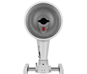 Anterior Segment Analysis
  Slit Lamp / Documentation
 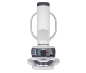 Myopia Management
 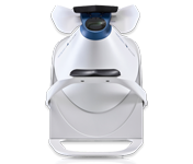 Perimetry
 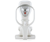 Tonometer
 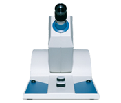 Visual Test Equipment
 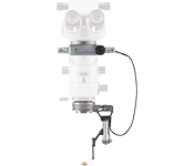 Vitreoretinal Surgery
  Manual Refraction
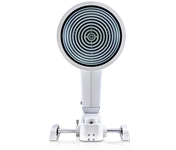
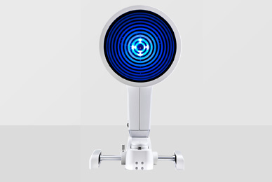
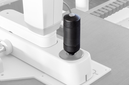
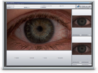
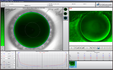
The OCULUS Keratograph 5M is an advanced corneal topographer with a built-in real keratometer and a colour camera optimized for external imaging. Unique features include examining the meibomian glands, non-invasive tear film break-up time and the tear meniscus height measurement and evaluating the lipid layer.

|
OCULUS Keratograph 5M |


With the Keratograph 5M, you can show your patients images they have never seen before. Gain patient trust by providing professional consultation during examinations and follow-ups.
Actively integrate the Keratograph into your consultation. Many easy to understand displays support you in communication and patient education. Use your Keratograph 5M as a marketing tool to make your services transparent!
The new joystick for the Keratograph 5M allows you to capture images and video sequences directly from the device. No need to use the mouse: your hand just stays on the joystick when you capture images or videos.
The new joystick communicates via a smart Bluetooth interface. And it does away with the need for cumbersome power cabling. The joystick switches to sleep mode function when not in use to extend battery life.
Switching to the new joystick is quick and simple. Watch the video below to see how easy it is!
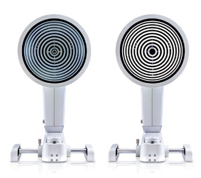
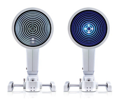
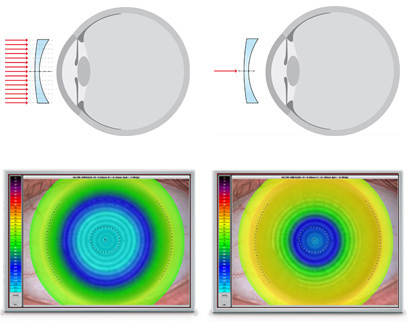
Thousands of measuring points are used to measure the whole surface of the cornea. A white ring illumination is used for this purpose. An infrared ring illumination is also provided for analysis of the tear film to prevent glare-related reflex secretion.
The perfect illumination has been integrated for every function of the Keratograph 5M: White diodes for the tear film dynamics, blue diodes for fluo-images, infrared diodes for Meibography.
 |
| Accuracy: | ± 0.1 D |
| Reproducibility | ± 0.1 D |
| Number of rings: | 22 |
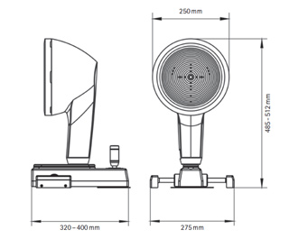
| Working distance: | 78 - 100 mm |
| Number of evaluated data points | 22 000 |
| Camera: | Digital CCD camera |
| Light source: | Placido illumination: White Placido illumination: Infrared 880 nm Fluorescein illumination: Blue 465 nmMeibography: Infrared 840 nmTear film dynamics: White Pupillometer illumination: Infrared 880 nm |
| Dimensions (W x D x H): | 275 x 320 - 400 x 485 - 512 mm |
| Weight | Measuring head: 3.2 kg (7.1 lbs) With base: 6.1 kg (13.5 lbs) |
| Max. power consumption | 18 W |
| Voltage | 90 - 264 V AC |
| Frequency | 47 - 63 Hz |
| Recommended computer specifications | CPU Intel® Core i5-7600, 8 GB memory, 1 TB, Windows® 10 Pro |
| Recommended screen resolution | 1920 x 1200 pixel |
In acc. with the Medical Device Directive 93/42/EEC

The OCULUS QM system is certified in accordance with ISO 13485 and (EU) 2017/745 (MDR).
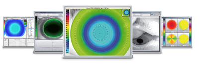
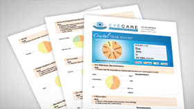
Find out the cause of dry eye disease quickly and reliably with the assistance of the new Crystal TEAR Report in the Keratograph 5M. Carry out a comprehensive analysis using the measurement results as a basis for your diagnosis. All results are summarized in a patient-friendly, neatly arranged printout, enabling you to combine documentation and patient education.
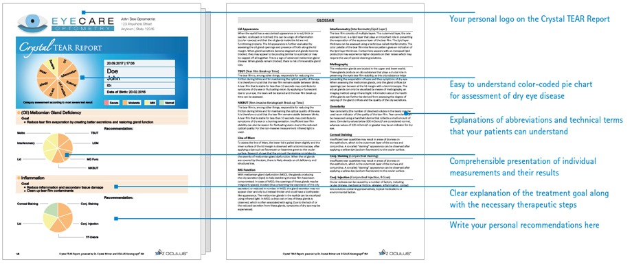
Benefit from all the advantages of the new Crystal TEAR Report in the Keratograph 5M: efficient screening, well-founded measurement results and greater patient loyalty.
With its preselection of recommendations and the option to manually adapt them, the Crystal TEAR Report allows you to draw up an effective treatment, according to the individual causes of
dry eye, in the shortest time. The result: a comprehensive dry eye analysis.
Up to 50 recordings can be carried out easily for documentation purposes. Readily understandable user tips and optimal auto-setting prior to each recording make it possible to delegate the entire measurement process. Despite the high degree of user guidance, you can directly delete faulty recordings as well as change settings and adapt the measurement sequence individually.
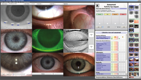
The entire course of diagnosis is documented, providing you with an ideal basis for your treatment decisions. All necessary recordings that are required to grade the individual's dry eye state, can be viewed at the same time. Depending on the grade you obtain, you can then determine a suitable treatment plan.
Additionally, you can enter the data from other tests, such as Osmolarity, Schirmer's Test, Phenol Red Thread Test, and more.
The Crystal TEAR Report surely and safely walks you through all the necessary assessment criteria that make up a comprehensive dry eye analysis. To complete the treatment plan, you can manually enter your dosage recommendations for the individual therapeutic measures on the printout. Making it an easy-to-grasp, optimal means of patient education.
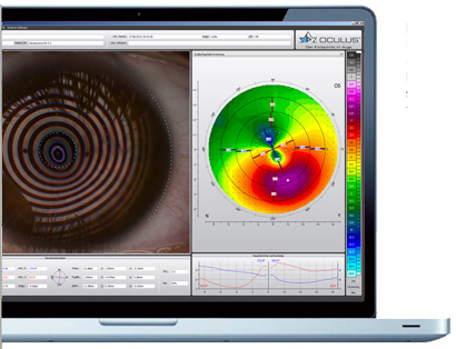
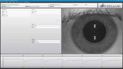
The tear film is assessed using either white or infrared illumination. The new high-resolution colour camera makes even the finest of structures visible. In addition to NIKBUT (Non-Invasive Keratograph Break-Up Time) and measurement of the meniscus tear, the TF-Scan can also make an assessment of the lipid layer and the tear film dynamics. Tear film analysis with the OCULUS Keratograph 5M is non-invasive and is conducted without any additional tools.
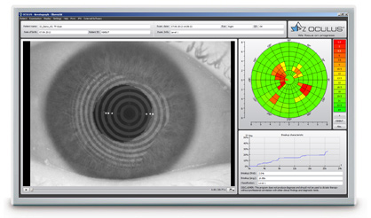
The tear film break-up time is measured non-invasively and fully automatically. The new infrared illumination is not visible to the human eye. This prevents glare during the examination. The TF-scan presents the results in a way that is easy for you and your customers to understand.
The height of the tear meniscus can be precisely measured with an integrated ruler and various magnification options, and its development along the edge of the bottom lid can be assessed. The results are saved to the patient file.
The interference colours of the lipid layer and their structure are made visible and can be recorded. The thickness of the lipid layer is assessed based on the structure and colour.
The video recording, with up to 32 frames per second, enables the observation of the tear film particle flow, from which conclusions regarding the viscosity of the tear film can be drawn.
Automatic classification of the bulbar redness. Conjunctival redness used to be assessed subjectively and depended on the examiner. The R-Scan is the first module that automatically and objectively documents and classifies the bulbar and limbal degree of redness. The R-Scan detects the blood vessels in the conjunctiva and evaluates the degree of redness. This automatic assessment spares you the time and effort of having to make manual comparisons using classification sheets.
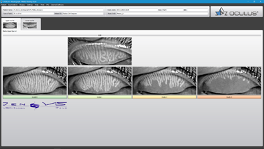
The multi-functionality of the Keratograph 5M easily and efficiently integrates difficult examinations such as meibography. The dysfunction of meibomian glands is the most frequent cause of dry eye. Morphological changes in the gland tissue are made visible using the Meibo-Scan and can be classified with the JENVIS Meibo Grading Scales.
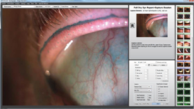
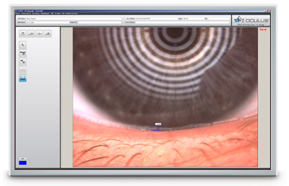
The Imaging Software is used to record video and image files. In addition to viewing the videos and single images by themselves, you can also compare the recordings with the simulated fluorescein-images of the measured eye.
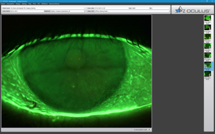
Result: Static fluorescein-images and videos can be recorded under slit lamp conditions!
The OxiMap® presents a colour map of the oxygen transmissibility of soft contact lenses based on the lens power, which is easy to understand – even for your customers!
An intact tear film and a good supply of oxygen to the cornea are an absolute necessity for wearing contact lenses comfortably. The OxiMap® presents a colour map of the oxygen transmissibility of soft contact lenses based on the lens power – which even your customers will understand!
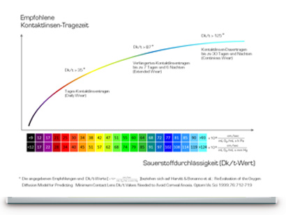
The oxygen transmissibility (Dk/t) depends on the material the contact lens is made of and on its thickness. In the past, only the manufacturers’ specifications about the oxygen transmissibility in the center of a -3,00 D contact lens were available. With the OxiMap®, you now get a graphic visualization of the Dk/t values over the whole area, based on the lens thickness.
The Dk/t values are colour-coded, whereby black represents an oxygen supply lower than that present in a closed eye. To preserve the integrity of the cornea when wearing contact lenses, minimum Dk/t values are recommended based on the length of time the contact lenses are to be worn. The OxiMap® colour-coding is based on these international recommendations.
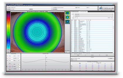
The OxiMap® is individually adapted based on the lens power and supports you in your consultation with the patient and helps you to choose the most suitable contact lens. New materials used for soft contact lenses provide excellent oxygen transmissibility.
OxiMap® was developed in close cooperation with JENVIS Research and the University of Applied Sciences in Jena.


With the OCULUS Newsletter you will receive free information about current events of our company.
Hot Topics
Product Categories
|
The OCULUS QM system is certified in accordance with ISO 13485 and (EU) 2017/745 (MDR). |